Tumors and Cancers of the Biliary Tract and Gallbladder
1. Gallbladder tumors and cancer
What type of tumors can be found in the gallbladder?
Both Benign and malignant tumors can occur in the gallbladder.
Benign lesions of the gallbladder are relatively common and include polyps such as cholesterol polyps. Approximately 5% of patients who have an ultrasound performed for abdominal pain will be found to have a gallbladder polyp. Only adenomatous polyps, though, are considered to have malignant potential.
Malignant tumors of the gallbladder are uncommon. Still, gallbladder cancer is the fifth most common gastrointestinal cancer. The most common malignant tumor of the gallbladder is adenocarcinoma.
What are the risk factors for Gallbladder Cancer?
Having a history of gallstones,
Having a history of chronic cholecystitis
Having other gallbladder conditions such as gallbladder polyps or chronic gallbladder infection,
Being female, and
Your risk increases with age.
Exposure to rubber or petroleum products may be a cause for adenocarcinoma
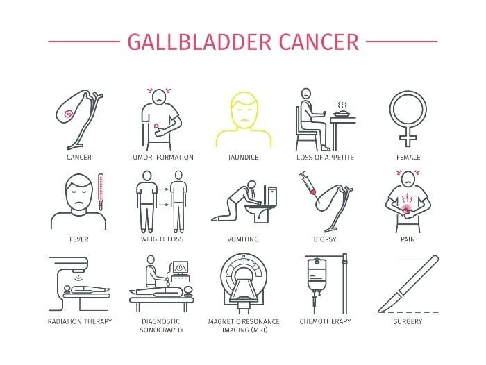
What are the symptoms of gallbladder cancer?
The symptoms of gallbladder cancer may be similar to those of acute cholecystitis (abdominal pain and fever), but there may also be no symptoms at all. If present, symptoms may include:
Abdominal pain or fullness, particularly in the right upper quadrant
Bloating
Fever
Unintentional weight loss
Nausea and/or vomiting
Night sweats
Back pain, especially pain that keeps you up at night
Jaundice (or yellowing of the skin and whites of the eyes) and
Abdominal mass (in the case of larger tumors or later disease)
How is a diagnosis of gallbladder cancer or other gallbladder tumors made?
Typically by imaging studies such as CT or ultrasound. Sometimes tumors will be found during endoscopic ultrasound or EUS.
How are Gallbladder Cancer and other Gallbladder Tumors Treated?
It depends on the type of tumor and whether or not it is malignant. Treatment options may include surgery, chemotherapy or radiation. In the case of small, benign polyps, surveillance may be acceptable based on patient preference and clinical guidelines.
2. Bile Duct Tumors
What Is Bile Duct Cancer (or cholangiosarcoma)?
Bile duct cancer is cancer that starts in the bile duct.
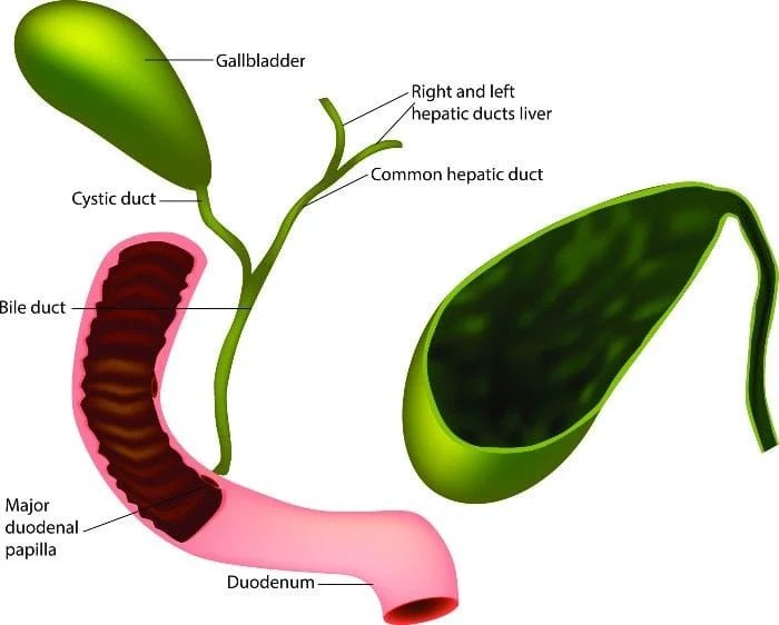
The bile ducts connect the liver and gallbladder to the small intestine. They transport bile from the liver and gallbladder to the small intestine, where bile aids in digestion.
What are the different types of bile duct cancers?
Cancer can start in any part of the bile duct system.
Malignant cancers that start in a duct inside the liver are called “Intrahepatic”
These cancers can sometimes be confused with liver or hepatocellular cancers. They are different though.
Malignant cancers that start in a duct outside the liver are called “Extrahepatic”
Extrahepatic bile duct cancers can be “perihilar” or “distal” depending upon the location of the tumor.
Perihilar cancers start at the hilum, where the left and right hepatic ducts have combined and are just leaving the liver
Distal bile duct cancers are found further down the bile duct, closer to the small intestine.
Nearly all bile duct cancers are adenocarcinomas, or cancers that start in the mucous cells that line the inside of the bile ducts.
Other less common types of bile duct cancers include sarcomas, lymphomas, and small cell cancers.
Not all bile duct tumors are malignant. Bile duct hamartomas and bile duct adenomas are examples of benign bile duct tumors.
What are the symptoms of bile duct cancer?
Most patients with bile duct tumors present with jaundice (yellowing of the skin or whites of the eyes). Other symptoms may include:
Itchy skin
Light colored or greasy/fatty stools
Dark colored urine
Abdominal pain or bloating
Loss of appetite
Unintentional weight loss
Back pain
Fever, chills, sweats
Nausea and vomiting.
What are the risk factors for bile duct cancer?
Having certain diseases of the liver or bile ducts such as Primary Sclerosing Cholangitis, liver flukes or parasites, bile duct stones, and/or Hepatitis B and C
Having Inflammatory Bowel Disease
Your risk increases with age
Hispanic Americans are at increased risk in the US. Worldwide, it is more common in Southeast Asia and China, largely because of the high rate of infection with liver flukes in these areas.
Being Obese
Having Non-alcoholic fatty liver disease (NAFLD)
A history of exposure to a radioactive substance called Thorotrast: a contrast agent used for x-rays until the 1950’s.
Having a history of Diabetes
Alcohol Abuse
A family history of Bile Duct Cancer
How are bile duct tumors diagnosed?
Because the tumors are generally small, standard imaging studies, such as ultrasound and CT may fail to show the lesion. MRCP, a special type of MRI, is sometimes used.
Other more invasive endoscopic approaches may be required to make a diagnosis.
How are bile duct cancers treated?
Treatment is dependent upon the site and extent of the lesion but may include surgery, chemotherapy and radiation.
3. Ampullary Tumors
What are Ampullary Tumors?
Ampullary tumors are malignant tumors found in the Ampulla of Vater, the last portion of the common bile duct as it passes into the duodenum (the first part of the small intestine). This is where all secretions from the pancreas, gallbladder, and liver enter the small intestine.
Tumors of the Ampulla of Vater can be benign (called adenomas) or malignant (referred to as ampullary carcinoma). Either can cause bile duct obstruction and be confused with gallbladder or pancreatic cancer.
Ampullary tumors are rare. Less than 1% of all GI cancers are Ampullary cancers.
What are the symptoms of Ampullary Cancer?
Symptoms may include:
Jaundice or yellowing of the skin and eyes
Loss of appetite
Weight loss
Abdominal pain
Back pain, especially back pain that keeps you up at night or is impossible to relieve
Itchy skin
Nausea, Vomiting, GI distress
Diarrhea
GI Bleeding
Pancreatitis
Pale, greasy, and fatty stools
How is Ampullary Cancer Diagnosed?
Diagnosis is typically made based on history, physical exam, lab tests and imaging studies to look for the presence of a tumor. Imaging studies may include ultrasound, CT or MRI. Sometimes endoscopic retrograde cholangiopancreatography (ERCP) or magnetic resonance cholangiopancreatography (MRCP) may be necessary. Abnormal lab findings typically reveal elevated Alkaline Phosphatase and bilirubin levels.
What are the treatment Options for Ampullary Cancer?
Treatment options may include surgery (specifically, a procedure called a Whipple procedure), chemotherapy, and radiation depending upon the extent of the disease.
Contact Us
Our Location
Find us on the map
Hours of Operation
Our Regular Schedule
Hunterdon Digestive Health Specialists
Monday:
8:00 am-6:00 pm
Tuesday:
8:00 am-7:00 pm
Wednesday:
8:00 am-5:00 pm
Thursday:
8:00 am-6:00 pm
Friday:
8:00 am-7:00 pm
Saturday:
*Contact office to confirm Saturday hours and availability*